Tissue perfusion pressure: MAP − Pcc, or Pcc − Pmsf?
Between the aorta and the vena cava, pressure falls in three steps: from arterial pressure down to the critical closing pressure, across the vascular waterfall to the mean systemic filling pressure, and from there to the right atrium. Two research groups have each named one of the first two steps "the pressure that drives tissue perfusion." They are not measuring the same thing.
Mechanism
Three pressure drops in series. Writing the whole circuit out:
MAP − CVP = (MAP − Pcc) + (Pcc − Pmsf) + (Pmsf − CVP)
The first term pushes blood through the arterioles and capillaries. The third is the gradient for venous return described by Guyton (see the Pmsf entry). The middle term is the vascular waterfall itself: the pressure that is "lost" at the point where vessels close, and which therefore does not contribute to flow on either side. Castro, Pinsky and colleagues drew this profile explicitly in their 2026 review (Castro et al., J Crit Care 2026, Fig. 2).
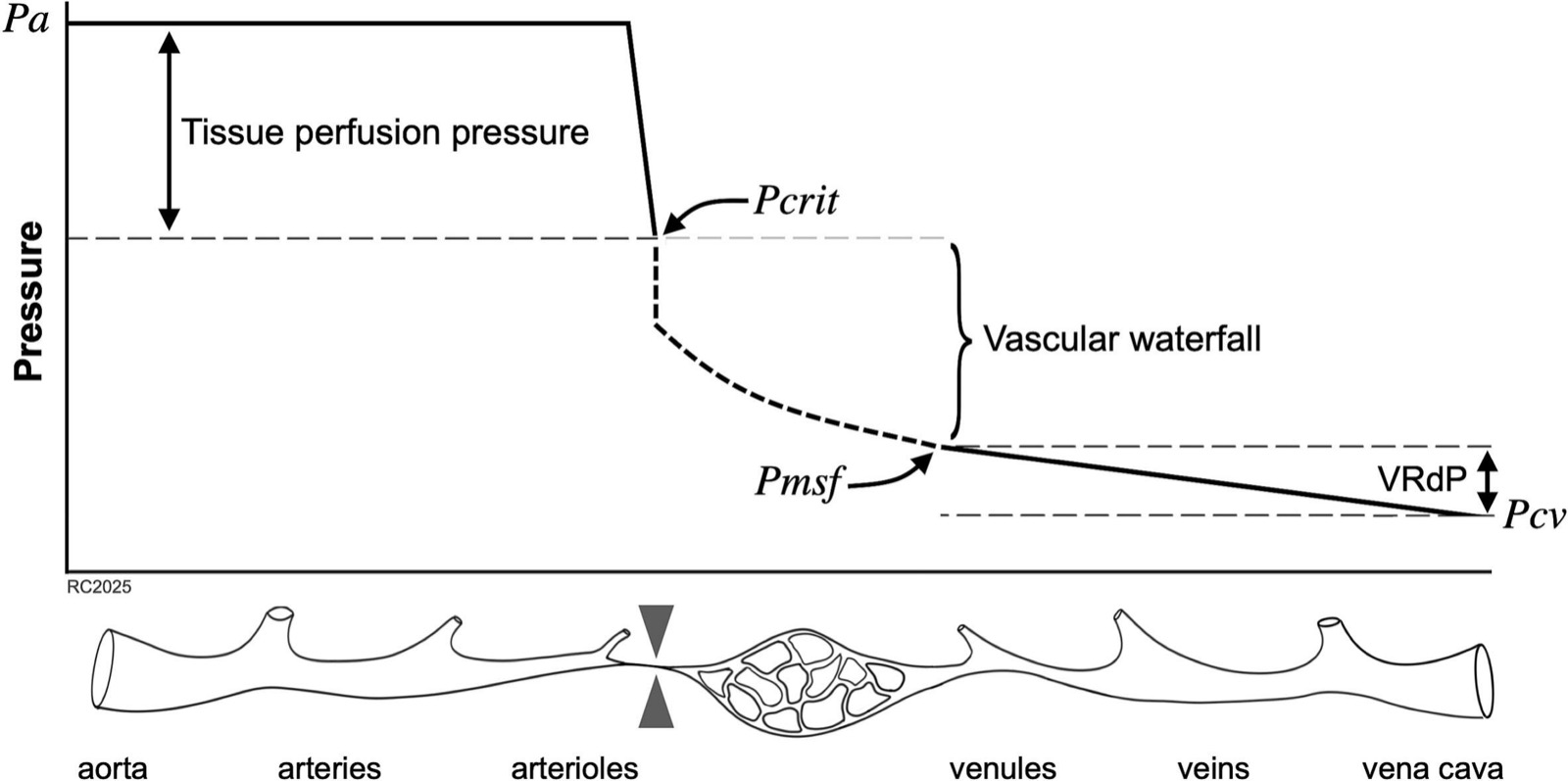
Why the waterfall disconnects the two sides. Permutt and Riley showed that once a collapsible vessel with tone is the limiting segment, flow depends only on the upstream pressure minus the closing pressure; the downstream pressure is irrelevant as long as it stays below the closing point (Permutt & Riley, J Appl Physiol 1963). The lung offers the familiar analogy: in West's zone 2, flow is set by arterial minus alveolar pressure, and venous pressure does not enter (West, Dollery & Naimark, J Appl Physiol 1964). In the systemic circulation the same logic separates an arterial pressure system from a venous one. Andrei and colleagues describe the waterfall as what "disconnects the arterial and the venous circulation in two pressure systems" (Andrei et al., Intensive Care Med Exp 2023).
The waterfall has a height, and the height moves. With high vascular tone, Pcc is high and the waterfall is tall. In vasoplegia, tone is lost, Pcc falls toward Pmsf, and the waterfall flattens, the two pressure systems reconnect. A vasopressor that restores tone raises Pcc and rebuilds the waterfall; whether it does so is a separate question from whether it raises MAP.
Two definitions of "perfusion pressure."
| Chandrasekhar and colleagues (2023–2026) | Andrei, Guinot and colleagues (2023–2026) | |
|---|---|---|
| Quantity | Tissue perfusion pressure, TPP = MAP − Pcc | Vascular waterfall, VW = Pcc − Pmsf |
| Measured how | Intercept of pulse pressure × heart rate against MAP; arterial side only | Inspiratory-hold manoeuvres; Pcc and Pmsf both measured |
| Described as | the pressure available to drive flow through tissue | "the pressure gradient driving microcirculatory flow" |
| Linked to | acute kidney injury, mortality, renal replacement | norepinephrine response, capillary refill time, pCO₂ gap |
The first is the step above the waterfall; the second is the waterfall itself. Both groups are working from the same physical model and cutting it at different places. Neither has been compared with the other in the same patients.
Evidence
The waterfall is low in vasoplegia, and norepinephrine does not always rebuild it. In thirty post-cardiac-surgery patients with vasoplegic hypotension unresponsive to fluid, baseline Pcc − Pmsf was only a few mmHg. Norepinephrine raised arterial pressure and total peripheral resistance in everyone. But in only 19 of 30 (63%) did the waterfall increase, from 3.5 to 43.6 mmHg, and in those patients cardiac index, capillary refill time and pCO₂ gap all improved. In the other 11, MAP rose just the same, the waterfall stayed flat, and the perfusion indices did not change. No baseline parameter predicted which group a patient would fall into (Andrei et al. 2023).
Capillary refill time tracks the waterfall, through its arterial end. In a larger cohort of 74 patients after cardiac surgery, treated according to phenotype with norepinephrine, fluid, or dobutamine, patients with prolonged capillary refill time had a lower waterfall (2.8 vs 18.3 mmHg), and changes in refill time correlated with changes in the waterfall (ρ = −0.40). The correlation was with ΔPcc (ρ = −0.42), not with ΔPmsf (ρ = −0.03) (Andrei et al., Ann Intensive Care 2026).
TPP adds information beyond MAP. In the cohort that introduced the measure, both TPP < 34 and MAP < 74 mmHg predicted outcome, but in external validation only TPP remained significant (Chandrasekhar et al., Nat Med 2023). After cardiac surgery, TPP < 38 mmHg was associated with acute kidney injury (Miles et al., J Thorac Cardiovasc Surg 2026). After heart transplantation, with cardiac output measured by thermodilution rather than estimated, TPP reclassified the risk of renal replacement therapy better than MAP (Wang C-C et al., J Heart Lung Transplant 2026).
Both ends of the waterfall in one cohort. Only one study has estimated Pcc and Pmsf together at scale: in 6,769 patients with sepsis, Pmsf was taken as the intercept of CVP against pulse pressure × heart rate, giving a waterfall gradient for each patient, with a threshold near 17 mmHg (Wang J-Y et al., Anesthesiology 2026).
Two cohorts that point in opposite directions, and the waterfall predicts both. After cardiopulmonary bypass, higher pre-bypass Pcc was associated with acute kidney injury (Ayers et al., J Cardiothorac Vasc Anesth 2025). In sepsis, lower Pcc was associated with mortality, with a U-shaped relationship (Wang J-Y et al. 2026). Read through the waterfall model these are the two ends of one curve: a low Pcc marks vasoplegia and a collapsed waterfall; a high Pcc marks excessive tone and a tall waterfall that the same MAP can no longer clear. Wang J-Y and colleagues report the U-shape directly.
What this means for a MAP target. Every finding above shares one implication. A MAP target cannot distinguish a patient whose Pcc is 25 mmHg from one whose Pcc is 55 mmHg, though at the same MAP their TPP differs by 30 mmHg. Nor can it distinguish a norepinephrine responder, in whom the waterfall was rebuilt, from a non-responder, in whom only MAP rose. The pressure that is targeted and the pressure that perfuses tissue are not the same number.
Open questions
Which gradient is "the" perfusion pressure has not been settled, because the two definitions have never been measured in the same patients. The pulse-pressure method yields only the arterial side and cannot compute Pcc − Pmsf; the inspiratory-hold method requires mechanical ventilation and a paused breath, and is impractical during surgery. Each method has settings it cannot reach.
The mechanism of norepinephrine non-response is unknown; the authors note that no baseline parameter predicted it (Andrei et al. 2023).
Estimates of Pcc from pulse pressure × heart rate have not been validated against stop-flow measurement; an accompanying editorial states that this accuracy "needs to be verified" (Pinsky, Anesthesiology 2026). The waterfall model itself assumes steady state and a fixed Pcc, and its authors label several proposed mechanisms as informed speculation (Castro et al. 2026).
No trial has used TPP or the waterfall gradient as a treatment target.